Detailed full-body images in one step
With the first total-body PET/CT in Germany, the Hannover Medical School is setting new standards in diagnostics, patient care and research - faster, more precise and with less radiation than ever before.
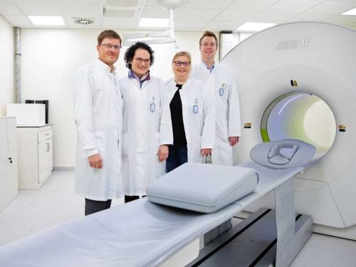
Top technology in MHH Nuclear Medicine: Prof. Dr. Frank Bengel, Prof. Dr. Sibylle Ziegler, Katja Henning, Medical Technologist for Radiology, and Dr. Philipp Rath are enthusiastic about the new device.
The Department of Nuclear Medicine at the Medical School Hannover (MHH) is the first institution in Germany and the fourth in the world to commission a so-called total-body PET/CT system with a field of view of around 150 centimeters. The innovative device can capture three-dimensional images of almost the entire human body in a single step. Lower Saxony's Science Minister Falko Mohrs came to the MHH campus for the official launch of the system. He explained that Lower Saxony is further expanding its pioneering role in nuclear medicine with the new total-body PET/CT: "The device, which is unique in Germany, will not only benefit patients at the MHH and the University Medical Center Göttingen thanks to its high-precision and fast imaging. It also opens up new possibilities in the research of certain diseases and their correlations. In addition, junior medical staff will benefit from this pioneering infrastructure, who will be able to work with state-of-the-art technology from the outset and secure this knowledge in Lower Saxony in the long term. The total-body PET/CT is therefore an investment in the future for Lower Saxony and far beyond." MHH President Prof. Dr. Denise Hilfiker-Kleiner underlined the importance of the new facility for the university: "With the introduction of total-body PET/CT, we are not only strengthening our research, but also enabling our patients to receive state-of-the-art diagnostics at the highest level."
Combined examination procedure
"The possibility of whole-body imaging has great potential for diagnostics, treatment and research. The device not only provides more precise images, it also works faster and with less radiation - these are significant advantages for patients," explained Clinic Director Prof. Dr. Frank Bengel. PET/CT technology is an important component of oncological, immunological, cardiological and neurological diagnostics. It combines two imaging techniques: positron emission tomography (PET) and computer tomography (CT). PET examinations are used to visualize molecular processes in the body. To make them visible, slightly radioactively labeled detection substances, also known as tracers, are used. One frequently used tracer is FDG, for example, a radioactively enriched type of sugar that helps to detect cancerous tumors. "Cancer cells consume more sugar. Due to their increased metabolic activity, we can identify tumors with the highest sensitivity," says Prof. Dr. Sibylle Ziegler, Head of PET Research at the Department of Nuclear Medicine. CT examinations, on the other hand, produce 3D cross-sectional images that show bones, organs and vessels in detail. In a combined PET/CT examination, both scans are carried out directly one after the other in the same unit. These images are then fused on the computer. "This brings together functional and anatomical information. Tumors or inflammations can be precisely localized," explains Professor Ziegler. The conventional PET/CT system at the Clinic for Nuclear Medicine can only capture a field of view of around 25 centimetres.
Images from the crown of the head to the thigh
The new total-body PET/CT system now takes this technology to a whole new level. Instead of the usual field of view of 25 centimetres, the new device offers a field of view of up to 150 centimetres. "We can create three-dimensional images of the human body from the crown of the head to the thigh with one image," says Professor Ziegler. "This means we can capture the molecular processes in almost the entire body at the same time. This makes it possible to view the body as a whole system, which goes beyond the usual view of individual organs." These overviews are very important, for example, when imaging metastases of an original tumor that are scattered throughout the body. Previously, nuclear medicine specialists had to take several small images one after the other and then combine them on the computer to create a large overview. Such an image took around 20 to 25 minutes. The new device can produce these large-scale images in less than a minute.
Faster and gentler
The shorter time in the "tube" is a great advantage for patients, for example if they cannot remain in one position for a long time due to pain or if they are small children who find it difficult to lie still for long periods of time. Patients also benefit from the fact that less radiation is required for total body PET/CT, making the examinations gentler. "The measurement technology is extremely sensitive and requires significantly lower amounts of administered radioactivity to achieve the required image quality," explains Professor Ziegler. Although less radiation is used, the images are higher-contrast and sharper than with conventional devices. The expert is particularly impressed by the fact that the images show much more detail. "The images have a resolution of up to three millimetres. That's fantastic when you consider that the previous standard was over four millimetres."
New opportunities for research
The ability to visualize molecular processes simultaneously in almost the entire body brings imaging diagnostics forward. It may now be possible to detect the success of a cancer therapy earlier and better and to adapt the therapy more precisely. The team at the Clinic for Nuclear Medicine will be able to examine significantly more patients per day with the new total-body PET/CT. However, the cutting-edge technology is not only used for clinical purposes. It also opens up completely new perspectives for researchers from numerous disciplines, for example when it comes to developing and using new tracers or new drugs.
Scientific opening symposium
A scientific opening symposium was held on January 23 to mark the commissioning. Prof. Dr. Simon Cherry from the University of California, Davis, a pioneer in the field of total-body PET/CT and a trailblazer in the technology, was the keynote speaker. The biomedical engineer considers the MHH to be one of the world's leading research centers for cardiovascular molecular imaging and is enthusiastic about the installation of the whole-body PET/CT scanner. "I am convinced that the combination of the best PET imaging technology and an outstanding team that understands the value of a systemic approach to disease research will lead to fundamental new insights with great clinical significance."
(Published: January 28, 2026)